


Skull tumor (osteoarthritis cranial) is a common, often chronic, disorder of the skeletal system that can cause serious, life-threatening complications if it develops. The disease has no cure. The word “Skull” in the name is not an exaggeration. Skulls are not bones.
The term describes a broad area of tissue that includes the intervertebral discs, the spinal cords, the muscles, the internal organs, the nerves, the tendons, the bursa, and the teeth.
Skull metastases are highly malignant bone tumors that are growing steadily in incidence among patients with osteoarthritis.
The study’s goals were to characterize the MR images, locations, and degree of invasion of the distal spinal sarcoma, evaluate patient outcomes and evaluate the potential long-term impact of surgical treatment options for the paravertebral subluxation associated with the skull base tumour.
This report presents the diagnostic results of a patient with a suspected or confirmed case of paravertebral subluxation. We discuss the treatment options and outcomes of this case.
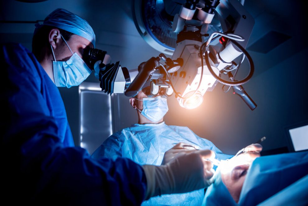
Brain Tumor and Skull Tumor Surgery Procedures
Localization of the tumor
A preoperative CT scan was requested. The biopsy findings included osteonecrosis (cracking) of the paravertebral bones, fibrosed-scalp pathology, paravertebral involvement and periarticular inflammation. The biopsy was completed after an initially lumpectomy and laminectomy.
Postoperative computed tomography scan and magnetic resonance imaging (MRA) were performed on the same day. After an initial plan was developed and written documentation was submitted, a preoperative MRI scan was performed. A preoperative MRI and magnetic resonance imaging scan was followed after a one-hour rest.
These two tests resulted in the following prognosis: a high-grade papillary cyst that required approximately five months of oral rest, no evidence of paravertebral tumor growth on the right ventricle or left the supranuclear region, no significant change in regional cerebral blood flow (CBIF) on the right or left sides, no significant changes in neurological function and no signs of infection.
Postoperatively, the radiologist performed localization of the tumor using an imaging system called a cerebrospinal fluid injection.
This imaging system is composed of a transducer that recognizes brain structures through its echo imaging sound waves and a computer. Other information such as the localization of the tumor, the amount of radiation needed to operate on it, and the surgical procedure’s clinical utility are also evaluated.
surgical resection of a tumour
Tumor Surgery is performed on an outpatient basis. Skull base tumour prognosis treated with standard surgical techniques have a high incidence of mortality. Tumor Surgery is performed under local anesthesia and generally has a high rate of complications.
Radiation therapy is very effective in the treatment of tumors of all types. Radiation therapy kills cancer cells and prevents their proliferation in healthy tissue. The type of radiation used depends on the type of tumor and its location. For instance, in brain tumors, brain radiation is usually delivered by x-rays, whereas it is less effective in treating non-cancerous paragangliomas and abscesses.
The need for surgical resection of a tumor of any type has increased in Toronto with the advent of a more significant number of critically ill children in the city. Many of these sick children have brain tumours or paragangliomas.
The surgical process can be delayed up to two years after the onset of symptoms in the case of non-evasive paragangliomas. It can also be deferred up to three years after the start of symptoms in the case of severely ill children. If resection is delayed longer, significant damage to normal healthy tissue is likely.
When the patient is stable after the Tumor Surgery, doctors usually use a stent in the skull base to keep the damaged portion closed while the diseased part heals. Patients with benign growth often require a single CT scan to assess their prognosis. If a significant improvement is noted, resection of the skull base can then follow. This is usually followed by radiation therapy to kill the remaining cancer cells.